Exciting new research from the AI Precision Health Institute at UH Cancer Center shows that deep learning can distinguish between mammograms of women who will develop breast cancer and those who will not.
X-rays were first used to detect breast cancer in 1913. Although breast imaging has evolved, the fundamental information that radiologists use to detect breast cancer has remained the same since 1913. Radiologists have always used morphology (assessing intensity of light, brightness, and shapes on images) to discern malignant breast cancer from benign lesions. Although radiologists have adopted new techniques such as CAD to improve diagnostic accuracy, they haven't analyzed new information on the image. Like human radiologists, CAD software only has morphology, texture, and image opacity available to make malignancy probability decisions.
Current techniques have such low sensitivity that millions of women are having unnecessary biopsies. In the United States only 29% of breast biopsies result in a breast cancer diagnosis. This means that 71% of breast biopsies could be avoided if radiologists could improve the accuracy in discerning what is benign and what is malign in breast tissue.
Many important advances in medicine have come from using AI to analyze medical images. In May of 2018, John Shepherd, PhD and colleagues published an important paper in Annals of Internal Medicine demonstrating that computers equalled radiologists in assessing breast cancer risk. This study also found that assessments by computer algorithm were more reproducible and less subjective than assessments by human radiologists.
Dr. Shepherd and his colleagues at the AI Precision Health Institute at the University of Hawaiʻi Cancer Center are using cutting edge computational resources and AI to facilitate research. They have collected mammography data dating back to 2009 from the major medical centers in Hawai‘i to form a mammography registry. This month they published two high profile papers detailing how they used AI to analyze new information to improve accuracy in detecting malignant breast cancer in medical images. The papers were published this month in Communications Medicine Nature and in Radiology Journal.
"The human eye can only see 256 levels of shades out of 65,000 on a mammogram. AI can see all 65,000 shades and compare thousands of variables relevant to cancer outcomes at the same time."
John A. Shepherd, PhD, study senior author, co-founder, AI Precision Health Institute
Dr. Shepherd, senior author of both studies, has been researching quantitative imaging for 30 years, and was one of the first to show that volumetric breast density measures are a stronger risk predictor than areal density measures. Dr. Shepherd is Interim Deputy Director and Chief Scientific Officer at University of Hawaiʻi Cancer Center, Professor of Population Sciences in the Pacific Program in Cancer Epidemiology, Director of the Shepherd Research Lab, and Director of the AI Precision Health Institute. Dr. Shepherd is known worldwide for his expertise in quantitative X-ray imaging using ML and the use of AI and DL to extract more cancer risk information from various forms of medical imaging including dual-energy X-ray absorptiometry, digital mammography, tomosynthesis, MRI, and 3D optical images. He has published over 200 peer reviewed papers and has been cited in other publications over 10,000 times. Dr. Shepherd and his colleagues are conducting biomedical imaging research to improve detection and monitoring of metabolic diseases and cancer risk.
Paper #1: Quantitative Mammography using AI and 3CB Imaging
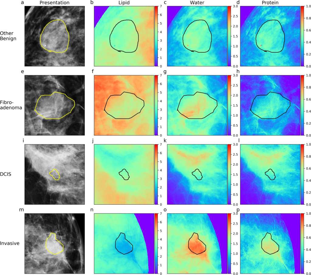
The first paper entitled Dual-energy three-compartment breast imaging for compositional biomarkers to improve detection of malignant lesions was published in Communications Medicine Naturein September 2021. In this paper Shepherd and his colleagues use a new technique called three-compartment breast imaging (3CB) that analyzes new information including lipid, water, and protein thickness to improve accuracy. The researchers confirmed that invasive breast lesions have unique compositional signatures when compared to other lesion types and demonstrates that compositional profiles of the breast combined with CAD predictions can improve the detection of malignant breast cancer.
In this study researchers used 660 breast images from 349 women. A neural network model was trained to predict probability of malignancy not lesion type. The researchers also looked at possible correlations between invasive cancers and patient hormone receptor status since it's hypothesized that cancers of different receptor type have unique compositional signatures due to utilization of exogenous fatty acids for sustained growth. The neural network model empirically demonstrated that adding compositional information improves classification of malignant and non-malignant lesions providing diagnostic value. The translational clinical benefit of this study is the increased confidence in the decision to perform a biopsy or not.
"Conventional methods of breast cancer risk assessment using clinical risk factors haven't been that effective. We thought that there was more in the image than just breast density that would be useful for assessing risk. In this study, invasive breast cancer tissues were found to exhibit lower lipid, higher protein and higher water content when compared to other non-invasive, non-cancerous breast tissues in which cancer was suspected."
John A. Shepherd, Ph.D., study senior author, co-founder, AI Precision Health Institute
AI has the exceptional ability to better classify breast cancer lesions using the lipid/water/protein signatures of the lesion calculated from the mammogram. Malignant lesions have unique biological and compositional characteristics so information including lipid, water, and protein thickness is important. To investigate, heat maps for lesions of each type were generated. Red indicates higher quantities of a given tissue component and quantities decrease as colors move towards violet. All lesion types, except DCIS, appear to have higher concentrations of protein and water relative to their background or surrounding tissue. The invasive lesions in particular appear to have a noticeably higher water signal.
Paper #2: Using Deep Learning To Predict Interval Cancer
The second paper entitled Deep Learning Predicts Interval and Screening-detected Cancer from Screening Mammograms: A Case-Case-Control Study in 6369 Womenwas published in Radiology Journal in September 2021. Researchers at the AI Precision Health Institute used AI to learn more from the mammograms than is possible using the human eyes. The objective is to be able to triage women by risk of breast cancer, to find cancers earlier, and to use resources where most needed.
This study was performed on over 25,000 mammograms. The mammograms obtained were from 6,369 women without breast cancer, 1,609 of whom developed screening-detected breast cancer, and 351 of whom developed interval invasive breast cancer. A deep learning model was trained to find details in the mammogram that might be linked to increased cancer risk and classify women into three groups: 1) those who did not develop cancer 2) those who developed screening-detected cancer 3) those who developed interval invasive cancer.
In this study deep learning models performed better than models using clinical risk factors in determining screening-detected cancer risk.This demonstrates that the use of deep learning to identify mammography features combined with traditional clinical risk factors increases the ability to predict risk of breast cancer. The models did not improve prediction of interval breast cancer based on breast density alone. This has implications for clinical practice because many management decisions today are guided by the use of breast density alone. While denser breasts on mammography are associated with a higher risk of cancer, there are other unknown factors that are hidden in mammograms that likely contribute to risk.
Interval cancers are cancers that are discovered within 12 months after normal screening mammograms. About 13% of breast cancers diagnosed in the US are interval cancers. Interval cancers usually have more aggressive tumor biology and are typically discovered at an advanced stage. It is therefore important to identify women who have a high risk for interval breast cancer and provide additional prevention strategies such as supplemental screenings.
"The results showed that the extra signal we're getting with AI provides a better risk estimate for screening-detected cancer. It helped us accomplish our goal of classifying women into low risk or high risk of screening-detected breast cancer. By ranking mammograms in terms of the probability of seeing cancer in the image, AI is going to be a powerful second reading tool to help categorize mammograms."
John A. Shepherd, Ph.D., study senior author, co-founder, AI Precision Health Institute
There are multiple AI algorithms approved by the FDA for detecting breast cancer. Three of these algorithms were compared in a recent external validation. The best of these algorithms was found to have a sensitivity of 82% in the detection of breast cancer, with a fixed specificity of 96.6%. The combination of this algorithm with a radiologist improved the sensitivity by 8%. With the risk algorithm presented in this study, negative mammograms would be further interrogated by AI to understand overall cancer risk and to make informed decisions on additional screening.
Future Studies Planned
Dr. Shepherd and his colleagues are planning to replicate this study using digital breast tomosynthesis and the women of Hawaiʻi that consist of a broad broad race and ethnicity profile. Most AI to date have been trained predominantly on white women. AI Precision Health Institute researchers also want to extend the work beyond cancer risk and look at women's risk of developing different grades of breast cancer, from least aggressive to most aggressive.
Margaretta Colangelo serves on the advisory board of the AI Precision Health Institute at the University of Hawaiʻi Cancer Center.
References
Dual-energy three-compartment breast imaging for compositional biomarkers to improve detection of malignant lesions, Communications Medicine Nature, September 2021 Collaborating with Dr. Shepherd were Lambert T. Leong, Serghei Malkov, Karen Drukker, Bethany L. Niell, Peter Sadowski, Thomas Wolfgruber, Heather I. Greenwood, Bonnie N. Joe, Karla Kerlikowske, Maryellen L. Giger. Author Affiliations: Department of Epidemiology and Population Sciences, University of Hawaii Cancer Center, Honolulu, Department Molecular Bioscience and Bioengineering, University of Hawaii at Manoa, Departments Epidemiology and Biostatistics, University of California San Francisco, Department of Diagnostic Imaging and Interventional Radiology, H. Lee Moffitt Cancer Center and Research Institute, Tampa, FL, Department of Information and Computer Science, University of Hawaii at Manoa, Department of Radiology and Biomedical Imaging, University of California San Francisco, Department of Medicine, University of California San Francisco, Department of Radiology, University of Chicago, Chicago
Deep Learning Predicts Interval and Screening-detected Cancer from Screening Mammograms: A Case-Case-Control Study in 6369 Women, Radiology Journal, September 2021 Collaborating with Dr. Shepherd were Xun Zhu, Ph.D., Thomas K. Wolfgruber, Ph.D., Lambert Leong, M.S., Matthew Jensen, B.S., Christopher Scott, M.S., Stacey Winham, Ph.D., Peter Sadowski, Ph.D., Celine Vachon, Ph.D., and Karla Kerlikowske, M.D. Author Affiliations: Department of Epidemiology, University of Hawaii Cancer Center, Department of Information and Computer Sciences, University of Hawaii at Manoa, Department of Health Sciences Research, Mayo Clinic, Rochester, Departments of Medicine and Epidemiology/Biostatistics, University of California San Francisco